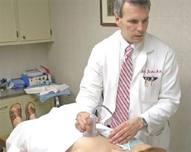
 Breast cancer is the most frequently diagnosed malignancy in women. Consequently, women should become actively involved in routine breast screening. This protocol includes monthly self-examinations, a yearly examination by a healthcare professional, and an annual mammogram beginning at age 40. Women with strong family histories of breast or gynecologic cancer, a personal history of cancer, or other risk factors, may require earlier and more frequent examinations. Any abnormality identified on physical exam or mammography will require additional investigation. Often, your physician will order tests such as ultrasound or MRI to further characterize these findings.
 When necessary, a tissue sample of the area in question will need to be obtained. If a lump can be felt in the breast, or if it can be seen on ultrasound, this area can easily be sampled in the office under ultrasound guidance using an automated core biopsy device. This requires no sedation or sutures, and is very accurate in acquiring tissue for the pathologist to examine. If the area in question can only be identified on mammography, then a sample must be taken either by a stereotatctic biopsy performed in the radiology department, or by a needle localized biopsy performed under sedation in the operating room. Your surgeon will discuss your options thoroughly, and assist you in making decisions regarding the type of biopsy best suited to your situation. The good news is that the majority of biopsies performed for suspicious areas in the breast prove to be benign (non-cancerous) lesions. The goal of breast screening is to identify breast cancer early, for early detection gives the best opportunity for cure.
If you are diagnosed with breast cancer, be encouraged that current treatment is very effective in producing an excellent opportunity for cure and improved survival. In most cases, the first step in treating breast cancer is the surgical removal of the cancer itself. In the past, the removal of the cancer necessitated removal of the entire breast along with the lymph nodes under the arm. This operation is called a modified radical mastectomy, and there are still conditions in which it will be the best option for a woman. Most women, however, are treated with a segmentectomy, also called a lumpectomy, in which the cancer is removed with a margin of normal breast tissue around it. This operation, coupled with radiation to the breast, gives the same opportunity for cure and survival as the mastectomy, but allows the woman to keep her breast. As a part of the surgical treatment of breast cancer, the lymph nodes under the arm must be evaluated for the presence of cancer cells, which may have spread from the breast tumor. The presence or absence of cancer cells in the lymph nodes is an important part of staging the cancer and in helping to direct additional treatment.
Again, in the past, most all of the lymph nodes under the arm were removed at the time of mastectomy, often leading to pain, limited shoulder movement, and the potential for lymphedema (swelling of the arm). Current techniques allow us to remove the few most important nodes under the arm through a small incision. These ‘most important’ nodes are called sentinel nodes, and their identification and removal is called a sentinel node biopsy. The presence or absence of cancer cells in the sentinel nodes accurately predicts the presence or absence of cancer cells in the remainder of the lymph nodes under the arm. Cancer cells identified in the sentinel nodes at the time of operation, generally necessitates the removal of the remainder of the nodes. Using these techniques of breast conserving surgery, the cancer can be removed and the nodes evaluated through two small incisions. This has become the ‘gold standard’ for the surgical treatment of breast cancer. After your operation, you will have an ongoing relationship with your surgeon, as part of your post-cancer screening protocol.
0 comments:
Post a Comment